|
|
Following modified from micro%2Ascope
|
Top | See original
| &pull 20q v5.145 20180528: Error 500 Can't connect to microscope.mbl.edu:80 (Name or service not known) http://microscope.mbl.edu/baypaul/microscope/lucidkeys/Euglena_key/html/species/sanguinea_des.htm |
|
Following modified from Department of Plant Biology Michigan State University
|
Top | See original
| &pull 20q v5.145 20180528: Error 301 Moved Permanently http://www.plantbiology.msu.edu/triemer/Euglena/euglenoid_cd/calliglena/calliglena_sp1.htm |
|
Following modified from Protist Information Server
|
Top | See original


Euglenophyceae
: Euglenales
Mastigophora: Euglenida
Euglena
sanguinea
Ehrenberg 1830
Syns:
E. viridis
var.
sanguinea
F. Stein,
E. haematodes
(Ehrenberg 1830) Lemmermann 1913
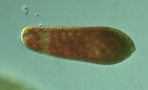
Species:
80-170
μm long,
25-45
μm wide; posterior bluntly rounded; flagellum about the body length; pellicle striated; chloroplasts
elongated
, parallel to the striae; haematochrome granules scattered in sun light and gathered in central area in darkness (Kudo, 1966). Body broadly fusiform;
90-120
μm long,
25-35
μm wide; anterior rounded, posterior tapered with a short process; chloroplasts tasselized and forming chromatophore bands, 12-16 in number; flagellum = body length (Photomicrographs of the Freshwater Algae, 1996).

E. sanguinea
106 x 28 μm
Saitama, 1999 |
E. sanguinea
cell μm
Saitama, 1999 |

E. sanguinea
144 x 22 μm
Mizumoto park
Tokyo, 2001 |

E. sanguinea
120 x 31 μm
Mizumoto park
Tokyo, 2001 |

E. sanguinea
165 x 27 μm
Ichikawa
Chiba, 2001 |

E. sanguinea
170 x 22 μm
Higashi-sano
Hakuba
Nagano, 2004 |

E. sanguinea
120 x 33 μm
Iwajuku
Gunma, 2001 |

E. sanguinea
110 x 27 μm
Minuma-hikawa P.
Saitama, 2002 |

Euglena sp.
156 μm
Tatebayashi
Gunma, 2001 |

E. sanguinea
147 x 32 μm
Kawashima
or Kawajima
Saitama, 2002 |

E. sanguinea
?
120 x 41 μm
Nanbata-shinden
Fujimi
Saitama, 2004 |

E. sanguinea
?
μm
Kashiwa Village m.p.
Kashiwa
Chiba, 2008

|
Please click on species name for more images.
Copyright
Protist Information Server
|
|
Following modified from Protist Information Server
|
Top | See original
Protist Movie Database
Euglenophyceae: Euglenales
Mastigophora: Euglenida: Euglenina
Euglena
sanguinea
Ehrenberg, 1830 (Syn.
E. haematodes
(Ehrenberg, 1830) Lemmermann, 1913):
Genus: Spindle, cylindrical or band-form; pellicle usually marked by longitudinal or spiral stirae; some with a thin pellicle highly plastic;
stigma
usually anterior;
chloroplasts
discoid, band-form, or fusiform;
two paramylum bodies
located on either side of nucleus,
rod-like to ovoid
in shape or
numerous and scattered
throughout; contractile vacuole near reservoir; cell division
longitudinal
(Kudo, 1966).
Species:
80-170
μm long,
25-45
μm wide; posterior bluntly rounded; flagellum about the body length; pellicle striated; chloroplasts
elongated
, parallel to the striae; haematochrome granules scattered in sun light and gathered in central area in darkness (Kudo, 1966). Body broadly fusiform;
90-120
μm long,
25-35
μm wide; anterior rounded, posterior tapered with a short process; chloroplasts tasselized and forming chromatophore bands, 12-16 in number; flagellum = body length (Photomicrographs of the Freshwater Algae, 1996).
Euglena sanguinea
, x 400, x 640, Saitama Pref., Japan, May-June 1999 by Y. Tsukii
Please click on images for viewing movies.
Copyright
1995-2007
Protist Information Server
|
Updated: 2024-05-02 12:24:41 gmt
|